Keywords
retina
neuroophthalmology
pseudotumor cerebri
virus
neuroophthalmology
pseudotumor cerebri
virus
How to Cite
Villegas, V. M., Monagas, M., Rivera, L., Santos, C., & Serrano, L. (2012). Visual Loss associated with Influenza A: a Case Report and Review of Literature. Puerto Rico Health Sciences Journal, 31(1). Retrieved from https://prhsj.rcm.upr.edu/index.php/prhsj/article/view/619
Abstract
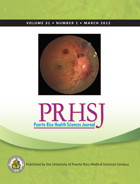
A 24-year-old female presenting with influenza A pneumonia and simultaneous visual loss was hospitalized. A complete ophthalmological examination was performed three weeks later. Best corrected visual acuity was 20/70 (right eye) and 20/30 (left eye). A dilated fundus exam revealed bilateral vitreous cells and marked bilateral optic nerve swelling with associated peripapillary hemorrhages. A submacular hemorrhage was seen in the right eye. Spinal tap opening pressure was 490 mmHg, with normal cerebrospinal fluid cell counts. Eight months after the initial clinical presentation, the patient was asymptomatic, with normal posterior poles and a best corrected visual acuity of 20/30 in the right eye and 20/25 in the left eye. Although papilledema can produce peripheral retinal hemorrhages secondary to extensive retinal venous congestion, the presence of bilateral vitritis and elevated influenza serum titers suggested that the patient might be suffering from influenza retinopathy. Vitreous polymerase chain reaction could potentially aid in the diagnosis of influenza retinopathy.
Authors who publish with this journal agree to the following terms:
a. Authors retain copyright and grant the journal right of first publication with the work simultaneously licensed under a Creative Commons Attribution License that allows others to share the work with an acknowledgement of the work's authorship and initial publication in this journal.
b. Authors are able to enter into separate, additional contractual arrangements for the non-exclusive distribution of the journal's published version of the work (e.g., post it to an institutional repository or publish it in a book), with an acknowledgement of its initial publication in this journal.
c. Authors are permitted and encouraged to post their work online (e.g., in institutional repositories or on their website) prior to and during the submission process, as it can lead to productive exchanges, as well as earlier and greater citation of published work (See The Effect of Open Access).